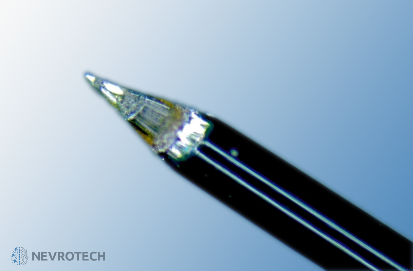
The Heptode Magic Pencil significantly improves spike sorting precision compared to the traditional microelectrodes or tetrodes. This performance is driven by a high-performance spatial recording characteristics, which allows for a more accurate identification of neuronal spikes.
Heptode Magic Pencil - HMP
The thin, Stainless Steel shaft of the probe contains 7 Platinum/Iridium electrodes, from which the middle one (tip electrode) is symmetrically surrounded by the 6 others. The Heptode Magic Pencil is characterized by low impedance value and remarkable signal-to-noise ratio (SNR). It is ideal for pre-clinical recordings.
Due to its shape, the Heptode Magic Pencil has the benefit of easy dural penetration resulting in minimal tissue trauma. For larger animals, such as primates a guide tube may be recommended for puncture.
The Heptode Magic Pencil significantly improves spike sorting precision compared to the traditional microelectrodes or tetrodes. This performance is driven by a high-performance spatial recording characteristics, which allows for a more accurate identification of neuronal spikes.
The thin, Stainless Steel shaft of the probe contains 7 Platinum/Iridium electrodes, from which the middle one (tip electrode) is symmetrically surrounded by the 6 others. The Heptode Magic Pencil is characterized by low impedance value and remarkable signal-to-noise ratio (SNR). It is ideal for pre-clinical recordings.
Due to its shape, the Heptode Magic Pencil has the benefit of easy dural penetration resulting in minimal tissue trauma. For larger animals, such as primates a guide tube may be recommended for puncture.
It may be possible to produce this probe beyond the listed parameters to facilitate the most ideal setup for your research needs. We can also build either capillaries (acute only) or fibers into the Heptode Magic Pencil for local drug delivery and optogenetical studies, respectively. Multiple connector types are available too for providing easy connection to your data acquisition system.
- 7 channels for single-unit or field potential recordings
- Improved spike sorting performance
- Stable impedance
- Excellent signal-to-noise ratio (SNR)
- Easy dural penetration
- Acute and chronic recordings
- Moderately customizable, durable design
- Capillary and fiber options
- Various connector types available
GENERAL
| Ideal method of use | Acute and chronic |
| Application method | In vivo or in vitro |
| Research phase | Pre-clinical |
| Subject | Small-, mid- and large-sized animals, such as primates |
| Shape | Symmetric |
| Linear | N |
| Steretrode | N |
| Tetrode | Y (heptode format is preferable) |
| Spacing if stereotrode or tetrode (µm) | – |
TIP
| Angle (°) | 30, 60 |
| Material | Epoxy, Stainless Steel, Platinum/Iridium (recording tip electrode) |
| Shape | Tapered (conical or sharpened) |
SHAFT
| Material | Stainless Steel |
| Diameter (µm) | 185 |
| Length (mm) | 10-200 (tip to the end of shaft) |
| Ferromagnetic | Y (non-MRI compatible) |
ELECTRODE SITES
| Material | Platinum/Iridium (placed concentrically) |
| Diameter (µm) | 15, 20, 25, 40 |
| Inter-electrode spacing (µm) | 50 |
| Number of electrode channels | 7 |
| Tip to 1st site distance (µm) | – |
CAPILLARY FLUID CHANNEL
| Applicable | Y (acute only) |
| Material | Glass |
| Outer diameter (µm) | 75 |
| Inner diameter (µm) | 50 |
FIBRE OPTICS
| Applicable | Y |
| Diameter (µm) | 75, 125 |
REINFORCEMENT TUBE
| Applicable | Y |
| Diameter (µm) | 250-640 |
| Length (mm) | 10-200 |
SILICONE DISK
| Silicone disk | N |
| Diameter (mm) | – |
| Thickness (mm) | – |
OTHER
| Connector types | Omnetics, Precidip (as per user’s request) |
| Lifespan | Reusable |
| Silicone cable between connector and probe (mm) | Chronic only |
| Special notes | It is important to test the capillary and/or the fiber optics before the first use. Cleaning is also recommended after each use. |
You may find downloadable content under Resources:
- Kóbor, Z. Petykó, I. Telkes, P.R. Martin, P. Buzás, ”Temporal properties of colour opponent receptive fields in the cat lateral geniculate nucleus,” 17 May 2017; 10.1111/ejn.13574
