Thumbtack Multielectrode Arrays have multiple recording sites in a configuration up to 32 electrode channels. They are designed to record electroencephalogram (EEG) signals from all layers of the cortex in parallel in vivo, and used in chronic applications in medium to large subjects, such as primates.
Thumbtack Multielectrode Arrays - TMA
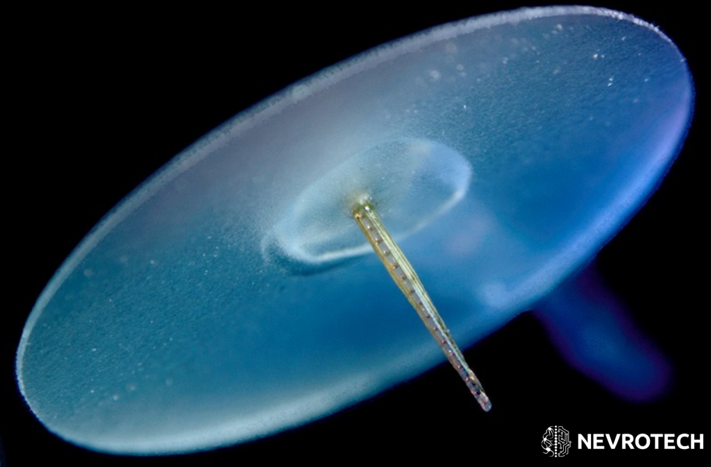
Thumbtack Multielectrode Arrays come with either a conical or sharpened tip to minimize trauma upon penetration. The recording sites are placed on the side of a symmetric epoxy cone with a choice of 4, 8, 16, 24 or 32 electrode channels with a maximum diameter of 500μm.
These Platinum/Iridium recording sites are available in 15, 20, 25 and 40μm diameter options. The smallest diameter of 15μm allows effective single-unit recording, and complemented with low impedance such probes have a better signal-to-noise ratio (SNR). Larger diameter probes are more suitable for field potential recordings.
Thumbtack Multielectrode Arrays have multiple recording sites in a configuration up to 32 electrode channels. They are designed to record electroencephalogram (EEG) signals from all layers of the cortex in parallel in vivo, and used in chronic applications in medium to large subjects, such as primates.
Thumbtack Multielectrode Arrays come with either a conical or sharpened tip to minimize trauma upon penetration. The recording sites are placed on the side of a symmetric epoxy cone with a choice of 4, 8, 16, 24 or 32 electrode channels with a maximum diameter of 500μm.
These Platinum/Iridium recording sites are available in 15, 20, 25 and 40μm diameter options. The smallest diameter of 15μm allows effective single-unit recording, and complemented with low impedance such probes have a better signal-to-noise ratio (SNR). Larger diameter probes are more suitable for field potential recordings.
In support of securing the Multielectrode Array in position and setting the depth of insertion, a very thin, round silicone disk is added perpendicularly to the end of the probe. Also, a silicone output lead with an integrated silk thread connects the probe to the connector.
Thumbtack Multielectrode Arrays are highly customizable and multiple connector types are available for providing easy connection to your data acquisition system. It may also be possible to produce these probes beyond the listed parameters to facilitate the most ideal setup for your research needs.
- Chronic recordings in medium to large animals
- Effective to record intra-cortical EEG signals
- Highly customizable design
- Conical tip to minimize trauma
- Platinum/Iridium sites (4-32 channels)
- Silicone disk to set the depth of insertion and to hold the TMA in place
- Various connector types available
GENERAL
| Ideal method of use | Chronic |
| Application method | In vivo |
| Research phase | Pre-clinical |
| Subject | Mid- and large-sized animals, such as primates |
| Shape | Asymmetric (tapering probe profile with silicone disk on top) |
| Linear | Y |
| Steretrode | N |
| Tetrode | Y |
| Spacing if stereotrode or tetrode (µm) | Min. 50 |
TIP
| Angle (°) | 15, 30, 60 |
| Material | Epoxy |
| Shape | Tapered (conical or sharpened) |
SHAFT
| Material | Epoxy |
| Diameter (µm) | 200-500 (varies based on the number of electrodes) |
| Length (mm) | 10-150 (tip to the end of shaft) |
| Ferromagnetic | N |
ELECTRODE SITES
| Material | Platinum/Iridium |
| Diameter (µm) | 15, 20, 25, 40 |
| Inter-electrode spacing (µm) | 50-500 (100 is typical) |
| Number of electrode channels | 4, 8, 16, 24, 32 |
| Tip to 1st site distance (µm) | 100-300 (combination of tip angle and probe diameter) |
CAPILLARY FLUID CHANNEL
| Applicable | N |
| Material | – |
| Outer diameter (µm) | – |
| Inner diameter (µm) | – |
FIBRE OPTICS
| Applicable | N |
| Diameter (µm) | – |
REINFORCEMENT TUBE
| Applicable | N |
| Diameter (µm) | – |
| Length (mm) | – |
SILICONE DISK
| Silicone disk | Y |
| Diameter (mm) | Min. 8 |
| Thickness (mm) | 0.15 |
OTHER
| Connector types | Omnetics, Precidip (as per user’s request) |
| Lifespan | Recording from 1 subject |
| Silicone cable between connector and probe (mm) | 15-300 |
| Special notes | – |
You may find downloadable content under Resources:
- Ulbert I, Halgren E, Heit G, Karmos G, “Multiple microelectrode-recording system for human intracortical applications,” 30 March 2001; J Neurosci Methods; 106(1):69–79.
- Cash SS, Halgren E, Dehghani N, Rossetti AO, Thesen T, Wang C, et al., “The human K-complex represents an isolated cortical down-state,” 22 May 2009; Science; 324(5930):1084–7.
- Keller CJ, Cash SS, Narayanan S, Wang C, Kuzniecky R, Carlson C, et al.; “Intracranial microprobe for evaluating neuro-hemodynamic coupling in unanesthetized human neocortex,” 15 May 2009, J Neurosci Methods; 179(2):208–18.
- Csercsa R, Dombovári B, Fabó D, Wittner L, Eross L, Entz L, Sólyom A, Rásonyi G, Szucs A, Kelemen A, Jakus R, Juhos V, Grand L, et al.; ” Laminar analysis of slow wave activity in humans,” September 2010; Brain; 133(9):2814-29.
-
Emília Tóth, Dániel Fabó, László Entz, István Ulbert, Loránd Erőss; “Intracranial neuronal ensemble recordings and analysis in epilepsy”; 15 February 2016; Journal of Neuroscience Methods; Volume 260, Pages 261-269